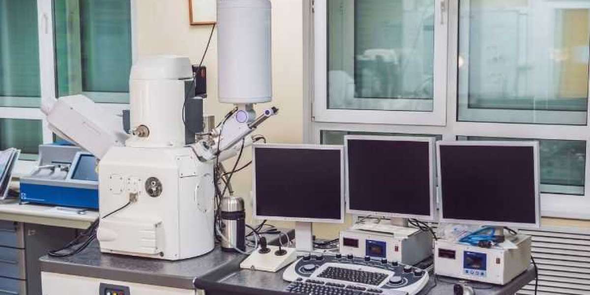
The global scanning electron microscope market size reached a value of around USD 4.49 billion in 2023. The market is further expected to grow in the forecast period of 2024-2032 at a CAGR of 8.30% to reach USD 9.24 billion by 2032 [1]. This phenomenal growth signifies the increasing dependence of various industries on a powerful tool – the Scanning Electron Microscope (SEM). But what exactly is an SEM, and how does it contribute to such diverse fields?
I. Introduction
A. Demystifying the SEM
A Scanning Electron Microscope (SEM) is a sophisticated imaging instrument that utilizes a focused beam of electrons to generate high-resolution images of a sample's surface. Unlike optical microscopes limited by light diffraction, SEMs achieve magnifications ranging from 20x to millions of times, allowing us to peer into the fascinating world of the unseen.
B. A Glimpse into the Power of SEMs
This blog post delves into the remarkable applications of SEMs across various industries. We'll explore how SEMs empower researchers, engineers, and scientists to unlock the secrets hidden within materials, cells, and even the tiniest particles.
C. Purpose of this Exploration
By understanding the diverse applications of SEMs, we gain a deeper appreciation for their role in scientific advancement and technological innovation. This knowledge not only sheds light on the intricate workings of our world but also paves the way for groundbreaking discoveries and solutions in the years to come.
II. Revolutionizing the Semiconductor Industry
The semiconductor industry forms the backbone of modern electronics. Here, SEMs play a critical role in ensuring the quality and functionality of integrated circuits (ICs) – the tiny chips that power our devices.
A. Inspecting the Microscopic World of Chips
SEMs are used to meticulously inspect the surface features of semiconductor devices at the nanometer scale. These examinations help identify defects such as cracks, misprints, and irregularities that could compromise the performance of the IC.
B. Nanometer Precision, Big Impact
The ability to detect defects at such a minute scale is crucial. Even the slightest imperfection can lead to malfunctions in the final product. By ensuring flawless construction, SEMs contribute significantly to the reliability and efficiency of modern electronics.
C. SEMs in Action: Examples from the Semiconductor World
- Analyzing the layout and connections within ICs
- Inspecting for shorts and opens in circuit pathways
- Examining the topography of transistors and other miniature components
III. Unveiling the Secrets of Materials Science and Engineering
From the development of new materials to understanding the properties of existing ones, SEMs are invaluable tools in materials science and engineering.
A. Peering Deep into the Material's Microstructure
SEMs allow researchers to study the microstructure of materials – the arrangement of grains, phases, and other microscopic features. This knowledge helps us understand how a material behaves and predict its performance under various conditions.
B. Composition, Phase, and Grain Structure – A Multifaceted Analysis
SEMs provide not only high-resolution images but also elemental analysis capabilities. This allows scientists to determine the composition of a material, identify different phases (regions with distinct properties), and analyze the size and distribution of grains within the microstructure.
C. SEMs: Illuminating the Path of Material Innovation
- Studying the microstructure of metals to optimize their strength and durability
- Analyzing the composition of alloys to develop new materials with tailored properties
- Investigating the microstructure of composite materials to understand their mechanical performance
IV. Bridging the Gap Between Biology and High-Resolution Imaging
The life sciences and biology fields have witnessed a paradigm shift with the introduction of SEMs. These powerful microscopes enable researchers to delve deeper into the intricate world of cells and organisms.
A. Imaging Biological Samples at Unprecedented Magnifications
SEMs allow us to image biological samples like cells, tissues, and microorganisms at magnifications exceeding those achievable with light microscopes. This detailed visualization provides invaluable insights into their structure, function, and interactions.
B. Unlocking the Secrets of Life: From Cells to Microbes
SEMs allow researchers to study the intricate details of cell morphology, including organelles, membranes, and surface features. Additionally, they provide insights into the structure and organization of tissues and help identify microorganisms responsible for infectious diseases.
C. SEMs: Pioneering Discoveries in the Biological Realm
- Studying the morphology of viruses and bacteria to understand their pathogenic mechanisms
- Analyzing the structure and function of specialized cells within tissues
- Investigating the interactions between cells and their environment
V. Unearthing the Earth's History: SEMs in Geology and Earth Sciences
Geologists and Earth scientists rely on SEMs to unlock the secrets hidden within rocks, minerals, and fossils.
A. Scrutinizing Geological Samples: A Window to the Past
SEMs provide high-resolution images and elemental analysis of geological samples, allowing researchers to study the composition, texture, and formation processes of rocks and minerals. This information sheds light on the Earth's geological history and the formation of various geological features.
B. Beyond the Surface: Unveiling the Secrets Within
SEMs allow researchers to analyze the internal structure of fossils, revealing details about the morphology and microstructure of ancient organisms. This information provides invaluable insights into past ecosystems and the evolution of life on Earth.
C. SEMs: Illuminating the Earth's Story
- Studying the composition and texture of rocks to understand their formation processes
- Analyzing the internal structure of fossils to reconstruct the morphology of ancient organisms
- Identifying trace elements in minerals to understand their origin and environmental conditions during formation


