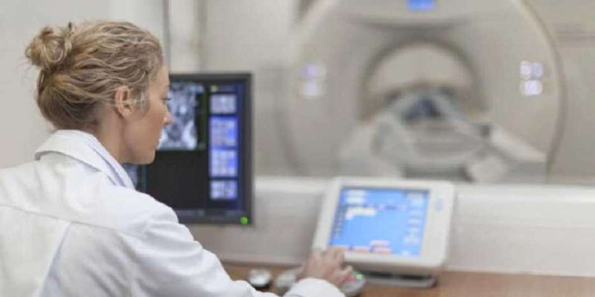
In the fast-evolving landscape of healthcare, diagnostic imaging plays a pivotal role in uncovering the mysteries of the human body. Among the myriad of imaging techniques available, ultrasound has emerged as a versatile and widely utilized tool. This blog aims to unravel the basics of ultrasound imaging, shedding light on its principles and applications in healthcare settings, with a particular focus on its significance in emergency rooms, often referred to as the "castle emergency room."
Understanding Ultrasound Imaging:
Ultrasound imaging, also known as sonography, is a non-invasive diagnostic technique that utilizes high-frequency sound waves to create real-time visualizations of internal structures within the body. Unlike other imaging modalities such as X-rays or CT scans, ultrasound does not involve ionizing radiation, making it a safer option for both patients and healthcare providers. Castle emergency room provide best Ultrasound imaging services.
Principles of Ultrasound:
Sound Wave Generation:
Ultrasound imaging relies on the transmission of sound waves through the body. A transducer, a device emitting these waves, is placed on the skin, and the echoes produced as the waves bounce off internal structures are captured.
Echo Detection:
The transducer not only emits sound waves but also detects the returning echoes. The time taken for the echoes to return and their intensity is used to create detailed images of organs, tissues, and blood flow.
Real-Time Imaging:
One of the key advantages of ultrasound is its ability to provide real-time imaging. This makes it invaluable for dynamic assessments, such as monitoring fetal development during pregnancy or visualizing blood flow in arteries and veins.
Applications of Ultrasound in Healthcare:
Obstetrics:
Ultrasound is widely used in monitoring fetal development, detecting anomalies, and ensuring a healthy pregnancy.
Abdominal Imaging:
It aids in assessing the liver, gallbladder, pancreas, and other abdominal organs for abnormalities or diseases.
Cardiovascular Imaging:
Doppler ultrasound is employed to evaluate blood flow and detect cardiovascular conditions such as blood clots or arterial stenosis.
Musculoskeletal Imaging:
Ultrasound helps assess soft tissues, muscles, and joints for injuries, inflammation, or structural abnormalities.
Ultrasound in Castle Emergency Rooms:
The term "castle emergency room" may evoke images of a fortress-like facility dedicated to handling critical medical situations promptly. In such high-pressure environments, ultrasound proves to be an indispensable tool.
Rapid Diagnosis:
Ultrasound allows for quick and efficient diagnosis of various conditions, facilitating prompt decision-making in emergency situations.
Bedside Imaging:
The portability of ultrasound machines enables healthcare providers to perform bedside imaging, reducing the need for patient transport and minimizing delays in critical care.
Trauma Assessment:
In emergency rooms, ultrasound is frequently used to assess trauma patients, providing crucial information about internal injuries, fluid accumulation, and the extent of damage.
Conclusion:
In the dynamic world of healthcare, ultrasound imaging stands out as a versatile and indispensable tool, providing valuable insights without the use of ionizing radiation. In castle emergency rooms, where every second counts, the rapid and reliable information provided by ultrasound can be a game-changer in the diagnosis and management of critical conditions. As technology continues to advance, ultrasound imaging will undoubtedly play an even more significant role in shaping the future of healthcare.


