
Breast Ultrasound Santa Fe
Breast Ultrasound Santa Fe health is a paramount concern for women, and medical advancements have made significant strides in the field of breast imaging. One such innovation is breast ultrasound, a non-invasive and safe diagnostic tool that plays a crucial role in early detection and accurate diagnoses.
What is Breast Ultrasound Santa Fe:
Breast Ultrasound Santa Fe In the vibrant city of Santa Fe, New Mexico, residents have access to cutting-edge healthcare facilities that offer state-of-the-art breast ultrasound services. In this article, we will explore the benefits of breast ultrasound in Santa Fe, shedding light on its importance in women's health.
I. Understanding Breast Ultrasound:
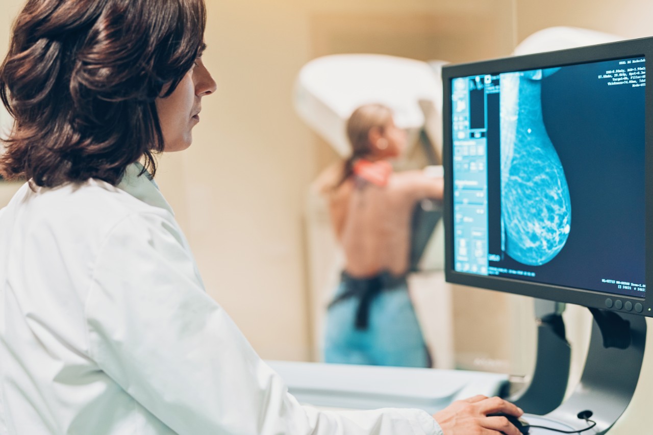
Breast ultrasound is a specialized imaging technique that uses sound waves to create detailed images of the breast tissue. Unlike mammography, which utilizes X-rays, ultrasound does not involve radiation, making it a safe option for both screening and diagnostic purposes. It is particularly useful for evaluating breast abnormalities found during physical examinations, mammograms, or other imaging studies.
II. Early Detection: Saving Lives:
Detecting Breast Cancer at Its Earliest Stage:
Breast ultrasound plays a crucial role in detecting breast cancer at its earliest stage when it is most treatable. It can identify suspicious areas, such as masses or lumps, that may not be visible on mammograms. By providing additional information about these areas, ultrasound helps doctors determine whether further testing or a biopsy is necessary.
Complementing Mammography:
Breast ultrasound is often used in conjunction with mammography to improve the accuracy of breast cancer detection. Mammograms are highly effective for most women, but they have limitations, especially for women with dense breast tissue. Ultrasound can help identify abnormalities that may be missed by mammography alone, ensuring comprehensive evaluation and reducing false-negative results.
III. Diagnosing Breast Abnormalities:
Characterizing Breast Lesions:
Breast ultrasound is instrumental in characterizing breast lesions, helping doctors differentiate between benign (non-cancerous) and malignant (cancerous) abnormalities. The technique provides valuable information about the size, shape, and composition of the lesion, aiding in the development of personalized treatment plans.
Guiding Needle Biopsies:
Ultrasound-guided needle biopsies are often used to collect tissue samples for further examination. By using ultrasound imaging to precisely target the abnormal area, doctors can ensure accurate sample collection, minimizing the need for invasive surgical procedures.
IV. Accessibility in Santa Fe:
The city of Santa Fe boasts a thriving healthcare industry that prioritizes women's health. Several reputable medical facilities in Santa Fe offer breast ultrasound services, providing residents with convenient access to this vital diagnostic tool. Patients can benefit from the expertise of highly skilled radiologists and technologists who are dedicated to delivering quality care and accurate diagnoses.
Conclusion:
Breast ultrasound has revolutionized the field of breast imaging, providing invaluable insights for early detection and improved diagnoses. In Santa Fe, women have access to state-of-the-art medical facilities offering breast ultrasound services. Through this non-invasive and radiation-free imaging technique, healthcare professionals can detect breast abnormalities that may not be visible on mammograms alone, leading to earlier interventions and better patient outcomes. As the importance of regular breast screenings continues to gain recognition, breast ultrasound remains a powerful ally in the fight against breast cancer, enhancing the overall quality of healthcare in Santa Fe and beyond.
Breast Ultrasound Santa Fe How Its Work?
Breast ultrasound is a diagnostic imaging technique that utilizes sound waves to create detailed images of the breast tissue. This non-invasive and radiation-free procedure plays a crucial role in the early detection and diagnosis of breast abnormalities, including cancerous and non-cancerous lesions. In Santa Fe, medical facilities equipped with advanced ultrasound technology provide this service to women, helping to ensure optimal breast health.
Here's how breast ultrasound works:
Preparation:
Before the ultrasound examination, you may be asked to undress from the waist up and put on a gown. To enhance the clarity of the images, it is important to remove any jewelry or clothing that may interfere with the procedure. You will be positioned comfortably on an examination table, usually lying on your back with your arm raised above your head.
Application of Gel:
A small amount of gel is applied to the skin of the breast, which helps in the transmission of sound waves and reduces friction between the skin and the ultrasound transducer. The gel is water-based and easily washes off after the examination.
Transducer Placement:
The ultrasound technologist, who is specially trained in performing the procedure, will gently press a handheld device called a transducer against the breast. The transducer emits high-frequency sound waves into the breast tissue, and these waves bounce back as echoes when they encounter different tissues and structures within the breast.
Image Formation:
The transducer picks up the echoes and sends them to a computer, which processes the information to generate real-time images of the breast. The images are displayed on a monitor, allowing the radiologist or technologist to interpret them and identify any abnormalities.
Scanning the Breast:
The ultrasound technologist will move the transducer across different areas of the breast to capture images from various angles. They may also apply gentle pressure to obtain optimal images and to evaluate the tissue's response. The examination may involve scanning the entire breast or focusing on specific areas of concern, depending on the reason for the ultrasound.
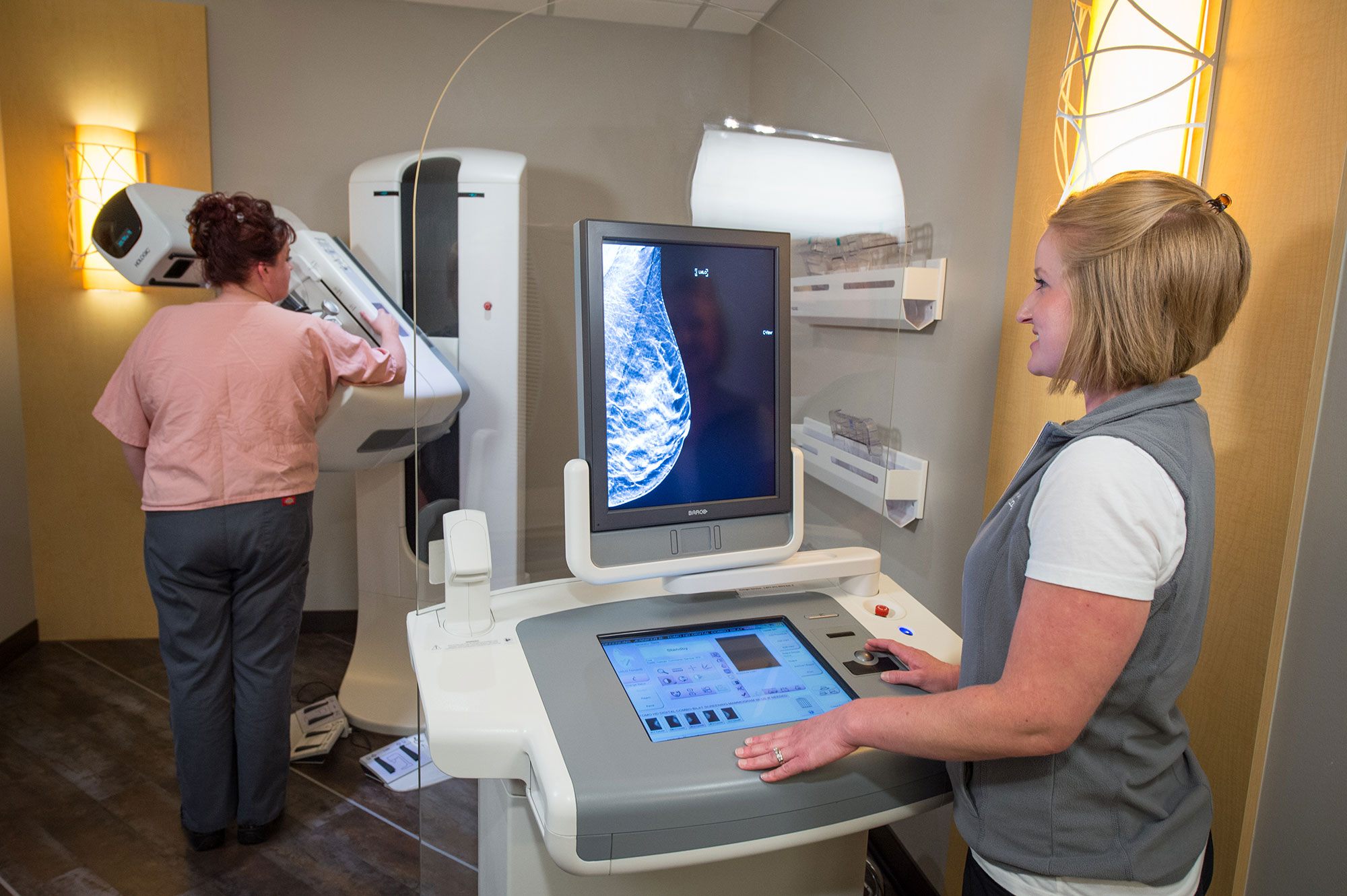
Real-time Imaging and Evaluation:
As the technologist performs the ultrasound, real-time images of the breast are continuously displayed on the monitor. The radiologist or technologist carefully examines these images to assess the shape, size, and composition of the breast tissue. They look for any abnormalities, such as masses, cysts, or other structural changes, and evaluate their characteristics.
Documentation and Reporting:
The findings from the breast ultrasound examination are documented in a report, which may include detailed descriptions of any abnormalities, measurements, and the radiologist's or technologist's impressions. This report serves as a valuable resource for further evaluation and treatment planning.
Breast ultrasound is generally painless and typically takes about 15-30 minutes to complete, depending on the complexity of the examination. After the procedure, you can resume your normal activities immediately, as there are no post-procedural restrictions or side effects.
If you want to get amazing benefits by using this link
Conclusion:
In Santa Fe, skilled radiologists and technologists are dedicated to performing breast ultrasounds with precision and accuracy. By harnessing the power of sound waves, breast ultrasound contributes to early detection, improved diagnoses, and ultimately, better patient outcomes in the field of breast health.